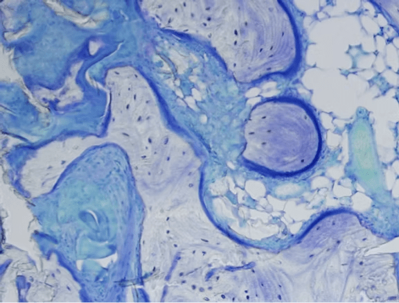
The critical role of proteoglycans in embryonic bone development is highlighted when genetic mutations are linked to skeletal disorders. Understanding the genetic basis of these disorders enhances our insight into critical design features of biomaterials for skeletal repair. One promising avenue is the use of carbohydrate-based scaffolds for tissue engineering. These materials, including Osteo-PTM BGS, structurally mimic GAGs and have shown potential for bone regeneration. For example, a novel hyper-crosslinked carbohydrate polymer demonstrated efficacy in repairing bone defects in vivo. To learn more, refer to “Hyper-Crosslinked Carbohydrate Polymer for Repair of Critical-Sized Bone Defects” (Koleva et al., 2019).

| Mutated Genes | Disease/ Disorders |
|---|---|
| PAPSS2, CHST3 | Skeletal dysplasia |
| IMPAD1, SLC35B2, CHST11 | Chondrodysplasia |
| CHST11 | Brachydactyly |
| CHSY1 | Temtamy preaxial brachydactyly syndrome |
| B4GALT7, CHST14, DSE | Ehlers-Danlos syndrome |
| SDC4 | Intervertebral Disc degeneration |
| XYLT1 | Desbuquois dysplasia type 2 |
| XYLT2 | Spondylo-ocular syndrome |
| FAM20B | Neonatal short limb dysplasia |
| EXT1 and EXT2 | Hereditary multiple exostosis |
| B3GAT3 | Larsen-like syndrome, Gerodermia osteodysplastica-like phenotype |
| EXTL3 | Skeletal dysplasia, Hereditary multiple exostosis |
| SLC35D1 | Schneckenbecken dysplasia |
| SLC26A2 | Chondrodysplasias, Achondrogenesis, Diastrophic dysplasia, Recessive multiple epiphyseal dysplasia |
| GORAB | Gerodermia osteodysplastica |
Proteoglycans are also key players in maintaining the structural integrity of the intervertebral discs, particularly through their role in collagen fibril organization and tissue hydration. In individuals with intervertebral disc disease, elevated levels of inflammatory cytokines such as tumor necrosis factor-α (TNF- α), interleukin (IL)-6, IL-1β can exacerbate disc degeneration. This process is mediated by the upregulation of the SDC4 gene which encodes for syndecan-4, a proteoglycan involved in matrix regulation. Activation of SDC4 triggers proteolytic cascades that lead to the disintegration of the disc extracellular matrix, primarily through enzymes like ADAMTS-5 and matrix metalloproteinases (Sao & Risbud, 2024).
| Gene | Normal Function | Dysfunction Due to Mutation | Disorder/Disease and Symptoms |
|---|---|---|---|
| XYLT1 | Xylosyltransferase , catalyzes the first step in GAG biosynthesis | Early chondrocyte maturation, ossification, leading to disproportionate dwarfism. | Desbuquois dysplasia type 2, Baratela–Scott syndrome |
| FAM20B | Xylose kinase for GAG chain extension | Immature, nonfunctional proteoglycans | Lethal neonatal short limb dysplasia, Skeletal phenotypes with reduced cartilage proteoglycans |
| B4GALT7 | Initiates GAG chain on the protein core | Proteoglycans lacking GAG chains, disorganized matrix | Progeroid Ehlers-Danlos syndrome |
| B3GAT3 | Adds glucuronic acid to the linker region | Reduced GAG chains, disorganized collagen | Larsen-like syndrome, Severe skeletal and cardiovascular anomalies |
| CHSY1 | Chondroitin sulfate synthase 1 | Disrupted bone morphogenesis, joint formation | Temtamy preaxial brachydactyly syndrome, Limb patterning defects |
| EXTL3 | N-acetylglucosaminyltransferase for GAG binding | Heparan sulfate synthesis | Skeletal dysplasia, Hereditary multiple exostosis |
| EXT1 EXT2 | Heparan sulfate polymerization in Golgi | Loss of function, skeletal develpment | Hereditary multiple exostosis |
| SLC35D1 | Golgi transporter for UDP sugars | Reduced GAG chain synthesis | Schneckenbecken dysplasia |
| SLC26A2 | Sulfate transporter | Reduced cartilage proteoglycan sulfation | Chondrodysplasias, Achondrogenesis, Diastrophic dysplasia |
| BGN | Biglycan, essential for bone and muscle | Severe osteopenia, collagen fibril abnormalities | Early onset of osteoporosis, disc degeneration |
| DCN | Decorin, regulates collagen fibril formation | Fragile skin and tendons | Irregular collagen fibrils, skin fragility |
| GORAB | Golgi protein recycling | Disrupted proteoglycan synthesis, collagen organization | Gerodermia osteodysplastica, Extreme bone fragility |
Chan, W. L., Steiner, M., Witkos, T., Egerer, J., Busse, B., Mizumoto, S., Pestka, J. M., Zhang, H., Hausser, I., Khayal, L. A., Ott, C.-E., Kolanczyk, M., Willie, B., Schinke, T., Paganini, C., Rossi, A., Sugahara, K., Amling, M., Knaus, P., … Kornak, U. (2018). Impaired proteoglycan glycosylation, elevated TGF-β signaling, and abnormal osteoblast differentiation as the basis for bone fragility in a mouse model for gerodermia osteodysplastica. PLOS Genetics, 14(3), e1007242. https://doi.org/10.1371/journal.pgen.1007242
Koleva, P. M., Keefer, J. H., Ayala, A. M., Lorenzo, I., Han, C. E., Pham, K., Ralston, S. E., Kim, K. D., & Lee, C. C. (2019). Hyper-Crosslinked Carbohydrate Polymer for Repair of Critical-Sized Bone Defects. BioResearch Open Access, 8(1), 111–120. https://doi.org/10.1089/biores.2019.0021
Paganini, C., Costantini, R., Superti‐Furga, A., & Rossi, A. (2019). Bone and connective tissue disorders caused by defects in glycosaminoglycan biosynthesis: A panoramic view. The FEBS Journal, 286(15), 3008–3032. https://doi.org/10.1111/febs.14984
Sao, K., & Risbud, M. V. (2024). Proteoglycan Dysfunction: A Common Link Between Intervertebral Disc Degeneration and Skeletal Dysplasia. Neurospine, 21(1), 162–178. https://doi.org/10.14245/ns.2347342.671
Taylan, F., & Mäkitie, O. (2016). Abnormal Proteoglycan Synthesis Due to Gene Defects Causes Skeletal Diseases with Overlapping Phenotypes. Hormone and Metabolic Research, 48(11), 745–754. https://doi.org/10.1055/s-0042-118706
Wen, J., Xiao, J., Rahdar, M., Choudhury, B. P., Cui, J., Taylor, G. S., Esko, J. D., & Dixon, J. E. (2014). Xylose phosphorylation functions as a molecular switch to regulate proteoglycan biosynthesis. Proceedings of the National Academy of Sciences, 111(44), 15723–15728.
© 2025 Molecular Matrix, Inc. All rights reserved.