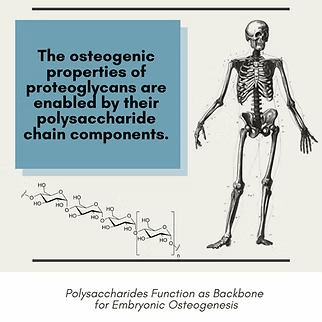
The ECM provides essential structural and biochemical support during embryonic osteogenesis. The structure of the ECM is composed of an interlocking mesh of proteins and long, unbranched polysaccharides called glycosaminoglycans (Raspanti et al. 2008). The majority of proteins are covalently attached to glycosaminoglycan chains, thereby forming proteoglycans. Proteoglycans are considered a class of proteins although they more closely resemble polysaccharides due to their 95% glycosaminoglycan composition. Types of proteoglycans are further classified on the basis of the glycosaminoglycan subtype they contain as opposed to their protein core. Proteoglycans have diverse functions in cartilage and bone, and display osteogenic properties contributing to embryonic osteogenesis (Iozzo and Murdoch 1996). Proteoglycans are expressed during both endochondral and intramembranous ossification and are concentrated in areas of active osteogenesis (Sugars et al. 2013). In cartilaginous tissues such as the early skeleton template, proteoglycans help form a stable matrix capable of withstanding compressive forces without losing structural integrity (Iozzo and Schafer 2015). Within the ECM, proteoglycans interact with other proteins, such as integrins, to facilitate cellular infiltration, cell to cell interaction, and cellular adhesion throughout the matrix. Certain proteoglycans have been found to bind and modulate the bioactivity of growth factors, including bone morphogenetic proteins, that are directly involved in the differentiation of chondrocytes and osteoblasts (Decarlo et al. 2012). Numerous studies show that proteoglycan deficiencies result in reduced osteoblast populations and activity, impaired bone formation, and failure to reach peak bone density at maturity compared to control organisms (Iozzo and Schaefer 2015).
References
Decarlo AA, Belousova M, Ellis AL, et al. Perlecan domain 1 recombinant proteoglycan augments BMP-2 activity and osteogenesis. BMC Biotechnol. 2012;12:60.
Gilbert SF. Developmental Biology. 6th edition. Sunderland (MA): Sinauer Associates; 2000. Osteogenesis: The Development of Bones.
Iozzo RV, Murdoch AD. Proteoglycans of the extracellular environment: clues from the gene and protein side offer novel perspectives in molecular diversity and function. FASEB J. 1996;10(5):598-614.
Iozzo RV, Schaefer L. Proteoglycan form and function: A comprehensive nomenclature of proteoglycans. Matrix Biol. 2015;42:11-55.
Kim T, See CW, Li X, Zhu D. Orthopedic implants and devices for bone fractures and defects: Past, present and perspective. Engineered Regeneration. 2020;1:6-18.
Koleva PM, Keefer JH, Ayala AM, et al. Hyper-Crosslinked Carbohydrate Polymer for Repair of Critical-Sized Bone Defects. Biores Open Access. 2019;8(1):111-120.
Raspanti M, Viola M, Forlino A, Tenni R, Gruppi C, Tira ME. Glycosaminoglycans show a specific periodic interaction with type I collagen fibrils. J Struct Biol. 2008;164(1):134-139.
Sugars RV, Olsson ML, Marchner S, Hultenby K, Wendel M. The glycosylation profile of osteoadherin alters during endochondral bone formation. Bone. 2013;53(2):459-467.
#osteogenesis #osteoconductive #polysaccharide #proteoglycan #HCCP #microenvironment
© 2025 Molecular Matrix, Inc. All rights reserved.