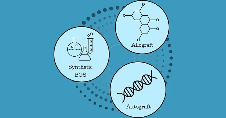
Osteoinductive – The graft must actively stimulate bone formation through recruitment of cells and molecules to the implant site.
Osteoconductive – The graft’s ability to support growth of new bone, allowing cells to adhere and multiply on the graft’s surface.
Osteogenic – The graft promotes the formation of new bone tissue.
Biocompatible – The graft material must be safe for implantation, minimizing the risk of infection or immune rejection.
Bioresorbable – The graft material should degrade at a controlled rate, as new bone is deposited.
Mechanical Strength & Porosity – The graft should provide adequate structural support while allowing for vascularization and tissue ingrowth, crucial for long-term bone health.
Growth Factor Delivery – The graft should act as a carrier for biological signals which enhance bone repair.
Non-toxic & Safe – The material must be free from antigenic, teratogenic, or carcinogenic effects, ensuring safety for patients (1-3).
Autografts are osteoconductive, osteoinductive, and have osteogenic properties
Autografts contain viable cells and natural growth factors crucial for healing, including bone morphogenetic proteins (BMP), fibroblast growth factors (FGF), vascular endothelial growth factors (VEGF), platelet-derived growth factors (PDGF), and insulin-growth factor 1 (IGF-1).
Autografts can be combined with other biologics such as bone marrow aspirate or platelet-rich plasma to reduce the amount of bone needed for the graft while enriching growth factor delivery (1-4)
Types of Allografts:
Cancellous allografts – Support structural bone repair
Cortical allografts – Provide denser load-bearing support
Demineralized Bone Matrix (DBM) – acid-extracted, removing minerals but preserving collagen and growth factors like BMPs, to support osteoconductive and osteoinductive action (2-4).
Osteoconductive – Allow new bone formation on BGS surface
Bioresorbable – Can be tuned to degrade as the bone heals, ensuring seamless integration
Mechanical Strength & Porosity – Support vascularization and tissue ingrowth
Some BGS are Osteoinductive & Osteogenic – Stimulating mesenchymal stem cell (MSC) proliferation
Forms of Synthetic BGS: Synthetic BGS are available in pellets, putty, powders, blocks, moldable, injectable, and 3D- printed bone scaffolds.
Calcium phosphate ceramics: tricalcium phosphate (TCP), biphasic calcium phosphate (BCP), hydroxyapatite (HA)
Bioactive glass: osteoconductive inorganic metallic oxides provide durability and strength
Calcium sulfate: temporary scaffold biomaterial
Biodegradable polymer biomaterials: polylactides (PLLA, PDLA), collagen, polyglycolide, and polycaprolactone (8)
Carbohydrate polymer-based scaffolds: such as our Osteo-P® BGS with biocompatibility, biodegradability which matches bone repair, and optimal porosity for osteoconductivity (9-11)
Composite biomaterials which blend ceramics and polymers to combine durability, biocompatibility, and improved bioactivity. Some formulations incorporate metals to enhance mechanical strength and promote osteogenesis (2-7)
[1] A. Bigham‐Sadegh, A. Oryan, Basic concepts regarding fracture healing and the current options and future directions in managing bone fractures, Int. Wound J. 12 (2015) 238–247. https://doi.org/10.1111/iwj.12231.
[2] C.E. Gillman, A.C. Jayasuriya, FDA-approved bone grafts and bone graft substitute devices in bone regeneration, Mater. Sci. Eng. C 130 (2021) 112466. https://doi.org/10.1016/j.msec.2021.112466.
[3] V.Al. Georgeanu, O. Gingu, I.V. Antoniac, H.O. Manolea, Current Options and Future Perspectives on Bone Graft and Biomaterials Substitutes for Bone Repair, from Clinical Needs to Advanced Biomaterials Research, Appl. Sci. 13 (2023) 8471. https://doi.org/10.3390/app13148471.
[4] J.W. Łuczak, M. Palusińska, D. Matak, D. Pietrzak, P. Nakielski, S. Lewicki, M. Grodzik, Ł. Szymański, The Future of Bone Repair: Emerging Technologies and Biomaterials in Bone Regeneration, Int. J. Mol. Sci. 25 (2024) 12766. https://doi.org/10.3390/ijms252312766.
[5] J.E. Inglis, A.M. Goodwin, S.N. Divi, W.K. Hsu, Advances in Synthetic Grafts in Spinal Fusion Surgery, Int. J. Spine Surg. 17 (2023) S18–S27. https://doi.org/10.14444/8557.
[6] J.R. Perez, D. Kouroupis, D.J. Li, T.M. Best, L. Kaplan, D. Correa, Tissue Engineering and Cell-Based Therapies for Fractures and Bone Defects, Front. Bioeng. Biotechnol. 6 (2018) 105. https://doi.org/10.3389/fbioe.2018.00105.
[7] M.A. Plantz, E.B. Gerlach, W.K. Hsu, Synthetic Bone Graft Materials in Spine Fusion: Current Evidence and Future Trends, Int. J. Spine Surg. 15 (2021) 104–112. https://doi.org/10.14444/8058.
[8] W.R. Moore, S.E. Graves, G.I. Bain, Synthetic Bone Graft Substitutes, ANZ J. Surg. (2001) 354–361.
[9] K.D. Kim, C.A. Batchelder, P. Koleva, A. Ghaffari-Rafi, T. Karnati, D. Goodrich, J. Castillo, C. Lee, In Vivo Performance of a Novel Hyper-Crosslinked Carbohydrate Polymer Bone Graft Substitute for Spinal Fusion, Bioengineering 12 (2025) 243. https://doi.org/10.3390/bioengineering12030243.
[10] P.M. Koleva, J.H. Keefer, A.M. Ayala, I. Lorenzo, C.E. Han, K. Pham, S.E. Ralston, K.D. Kim, C.C. Lee, Hyper-Crosslinked Carbohydrate Polymer for Repair of Critical-Sized Bone Defects, BioResearch Open Access 8 (2019) 111–120. https://doi.org/10.1089/biores.2019.0021.
[11] S. Michael A, L. Charles C, Bridging and Repair of First Metatarsal Fracture with Chronic Pseudoarthrosis Following Multiple Surgical Interventions Using a Minimally Invasive Approach, Open J. Orthop. Rheumatol. 10 (2025) 001–004. https://doi.org/10.17352/ojor.000050.
© 2025 Molecular Matrix, Inc. All rights reserved.